We headed east today in search of a dive spot protected from the swell, with hopes that the wind would die down. Good fortune, as conditions improved, and we were able to explore both the protected southeastern and exposed northwestern ends of a small submerged platform located at the confluence of two deepwater channels.
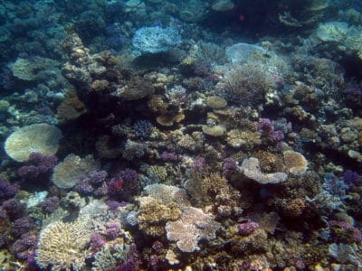
Farasan Banks Acropora garden.
Besides two of the most breathtaking wall dives we have surveyed in the middle portion of the Farasan Banks, we were greeted by a small bryde whale (pronounced broo-dess, Balaenoptera edeni), a type of minke whale commonly seen in the southern Red Sea. He was swimming slowly along the channel, and must have come a bit close to the reef, breaching twice for a brief moment each, before diving to the depths. We first spotted him from the boat, during our surface interval. Within seconds of the first siting, the team donned mask, snorkel and fins and jumped off the boat, hoping to get a photograph. No such luck, maybe next time. These whales are found in coastal waters of the Indian and western Pacific oceans and Red Sea, where they feed on pelagic fish crustaceans and cephalopods. Their populations were seriously knocked back as a result of historical whaling practices; up to about 80,000 thought to be remaining in the year 2000.
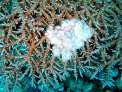
A growth anomaly on a table acroporid (Acropora pharensis).
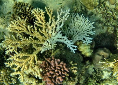
The leeward side had the richest reef flat community we’ve seen to date, with thickets of acroporids in every growth form imaginable. We saw tree-like (arborescent), upright staghorn-like, bottlebrush, finger-like (digitate), bushy, clumping and plate-like tables. Colonies ranging from a few centimeters to several meters in diameter, and stands that extended for 10s of meter. They were every color of the rainbow, ranging from purples, to red and deep blue, as well as yellow, green and light brown, and including many species we have not yet seen in the Farasan Banks.
Other corals, such as the fat-branched, purple colored Pocillopora, small massive lobes of Porites were intermixed, but all corals in this shallow environment (1-3 m depth) were not excessively tall, generally less than 1 m in height, probably to avoid heavy wave action and periodic low tides. At the edge of the reef flat, the reef dropped abruptly to a gently sloping terrace at 10-15 m depth, before plummeting vertically to the depths.
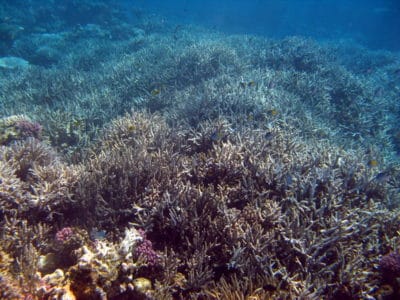
A thicket of Acropora formosa.
The terrace had a diverse assemblage of small massive corals, with many larger acroporids that towered above the community, forming plates or tables, often a few meters in diameter.Unexpectedly, my dive buddy Bernhard pointed at one of these in the distance with an unusual tumor-like growth anomaly. While these are generally quite common, worldwide, being one of the first recognized diseases (identified by Dr. Squire in 1965), we had not seen many colonies with the growth anomalies at other sites in the Farasan Banks yet.
I slowly kept swimming, closely examining each of the table acroporids. What I noticed were a number of dead colonies, and many that were partially dead, and colonized by algae.
I also spotted several with a disease known as white syndrome, which has been recently reported from throughout the Indo-Pacific including Australia, Palau, the Philippines and several remote U.S. Pacific insular areas. It turns out this condition was extremely common among deeper water (10-22 m depth) table acroporids at this reef. I observed over 25 colonies with recent tissue loss in an area several hundred meters wide.
The disease exhibits a very characteristic pattern of tissue loss that spreads across the colony like a forest fire, starting at one point and progressively radiating outward. Areas first affected are colonized by algae, and over time colonies exhibit a characteristic progression.
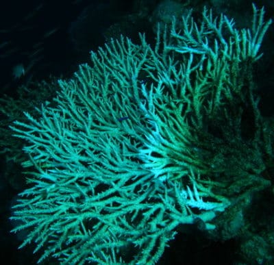
A table Acropora with white syndrome.
Closest to the living tissue is a band of white, recently denuded skeleton. This grades into a greenish colored band (filamentous algae and diatoms) with clearly visible coral skeletal elements, and eventually brownish or reddish as crustose coralline algae and other alage and invertebrates colonize the skeleton.
Its easy to mistake a disease for other natural stressors, such as predation by the crown of thorns sea star, Acanthaster (COTS), which can remove tissue in a similar manner.
Also these starfish usually feed at night, and retreat to the base of the coral, or a crevice in the reef during the day, to avoid being eaten themselves. I carefully searched around each colony and the adjacent areas, but found none.
A colony of Acropora austera eaten by Acanthaster, and a second colony partially eaten. The COTS is on the right side.
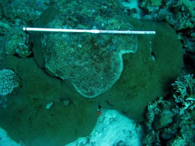
A slowly spreading disease (white syndrome) on a star coral (Diploastrea).
The crown of thorns starfish was present on this reef, though, in very low numbers. In the shallows, there were also a number of dead colonies and some with recent tissue loss. At the base of one of these corals, I found the culprit, a small starfish tucked in a hole, out of site to predators. I also found one on the windward side, again in the reef flat, and once again, there were several corals that the starfish had eaten very recently.
While the white syndrome seemed fairly common on this reef, and it affected a few other corals such as a large massive colony of a type of star coral (Diploastrea), there were hundreds of juvenile corals (10-20 cm) in perfect health, and lots and lots of newly settled corals at all depths, suggesting that those larger, old colonies that were killed by white syndrome were being replaced by many new colonies.
