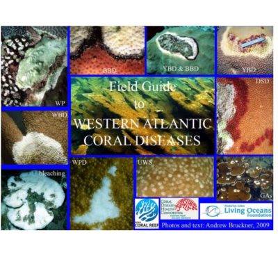
Field Guide to Western Atlantic Coral Diseases
(2010)
Please find an excerpt of the full PDF below
Field Guide to Western Atlantic Coral Diseases
 This Field Guide to Western Atlantic Coral Diseases uses standardized nomenclature and diagnostic criteria for coral diseases developed by the Coral Disease and Health Consortium (CDHC). This involves a three-tiered approach to naming diseases and a decision tree that would help unify disease names. Through application of a series of steps, a researcher can determine a common field name for a disease which can be further refined based on a morphologic diagnosis and an etiologic diagnosis. This is based on techniques applied to the study of other terrestrial and marine animal diseases that include 1) detection of disease, 2) description of the morphologic changes in the host associated with the disease, 3) evaluation of morphologic changes in the host as disease progresses (e.g., pathogenesis), determining the cause (e.g., etiology), and physiologic changes in the host as the disease progresses (e.g., pathophysiology). These underwater cards help a diver complete a level 1 diagnosis.
Level 1:
Categorization of obvious gross field signs directly visible to the diver without magnification or sampling. The first step involves the identification of four easily observed descriptive categories – presence of color change, tissue loss, skeletal damage and irregular growth. The observer also records other gross visible signs such as the location and distribution of the lesion, condition of the affected tissue, and the extent of tissue loss. Based on an analysis of these characters, it is possible to identify a common field name to describe the disease in the particular coral and apply the same terminology to other corals (regardless of species) with similar gross visible signs. This approach is used primarily for rapid assessment and routine monitoring.
Level II:
Detailed examination of affected and unaffected tissue taken from the diseased coral (and compared to representative presumed healthy corals) using histology. This approach can help identify the presence of microorganisms and describe morphological changes to the tissue. This could result in a morphologic diagnosis.
Level III:
A detailed set of field and laboratory tests to identify and confirm the presence of proposed causative agent (s), toxin, or other factor responsible for the manifestation of the disease. Microbial diseases may be determined according to Koch’s postulate – where a presumed pathogen is isolated, grown in pure culture, identified, and used to infect a presumably healthy host. If the disease signs appear, and the presence of the presumed pathogen is confirmed, and an etiologic diagnosis can be assigned.