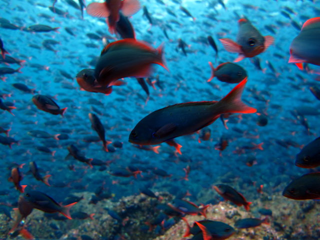
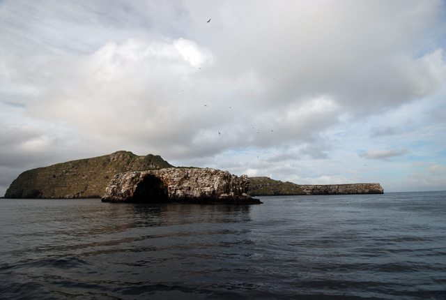
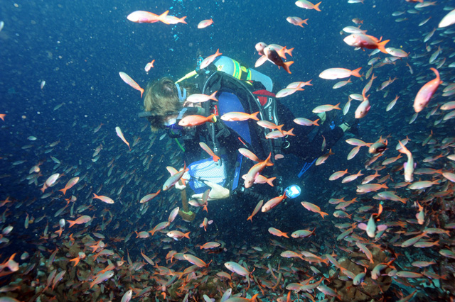
When we entered the water for our first research dive at Wolf island, the first thing we saw were thousands upon thousands of fish. The fish were so numerous it made seeing the bottom difficult. Underneath the clouds of fish the dominant species of coral was Porites lobata, a lobed pore coral. This species forms massive, helmet-shaped or hemispherical colonies that can exceed 3 m in diameter and 2 m high.
This long-lived coral is slow growing (1-2 cm/year), but is generally more resistant to diseases and bleaching than other corals. It recovered fairly quickly from two severe El Niño events (1982-1983 and 1997-1998) and it is the dominant species around both Wolf and Darwin Islands.
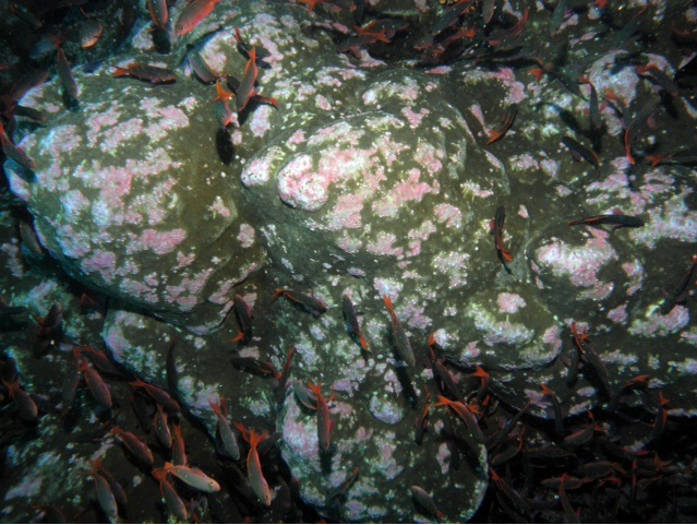
This coral is a favorite of coral eating fish, and the colonies we saw were typically covered with bite marks. In addition to numerous small excavations from fishes, we noted that a high proportion of the colonies have discolored patche, spots, and bands that range in color from pink to blue. Scientists have seen these kind of pink spots discoloration in other areas.
In Hawaii, the genus Porites is frequently infected with trematodes. In response to the infection, the coral develops a small nodule, pink in color, to encase the parasite. These pink spots are selectively preyed upon by butterflyfish, which serve as a host for one life stage of the trematode.
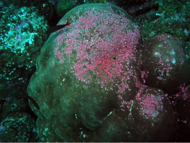
Recently, a new disease called pink line disease emerged on Porites colonies from the Arabian Gulf. There is considerable dispute of the origin of these pink spots and bands, as they can also be caused by any irritation caused by a fish bite, algal abrasion, barnacles, polychaete tube, and other invertebrates. One thought is that this is a stress response of the coral, and the discolored area may actually serve as a barrier, protecting unaffected parts of the colony.
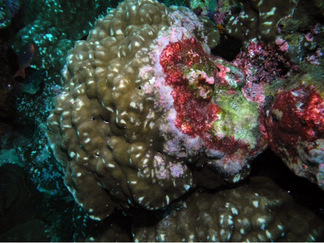
We observed an unusually high prevalence of pink spots and bands during our surveys at Darwin and Wolf Islands, much higher than that reported in the past. These have a wide variation in appearance and may be associated with obvious injuries and fish bites. We also saw the discoloration forming more of a characteristic band-like pattern that is advancing across the coral, denuding tissue behind it. To better understand the causes and effects of these pink spots on the colony, Dr. Andrew Bruckner collected tissue samples, which he will examine using histology.
(Photos by: 1 Dr. Brian Beck, 2 Alison Barrat, 3-5 Dr. Andy Bruckner, 6 Dr. Brian Beck)
To follow along and see more photos, please visit us on Facebook! You can also follow the expedition on our Global Reef Expedition page, where there is more information about our research and team members.


